

AI-assisted dermatology workflows with advanced imaging and intelligent lesion tracking

Dermatology

Advanced skin imaging, AI lesion tracking, for modern dermatology practices

07:42 — a teledermatology intake lands.

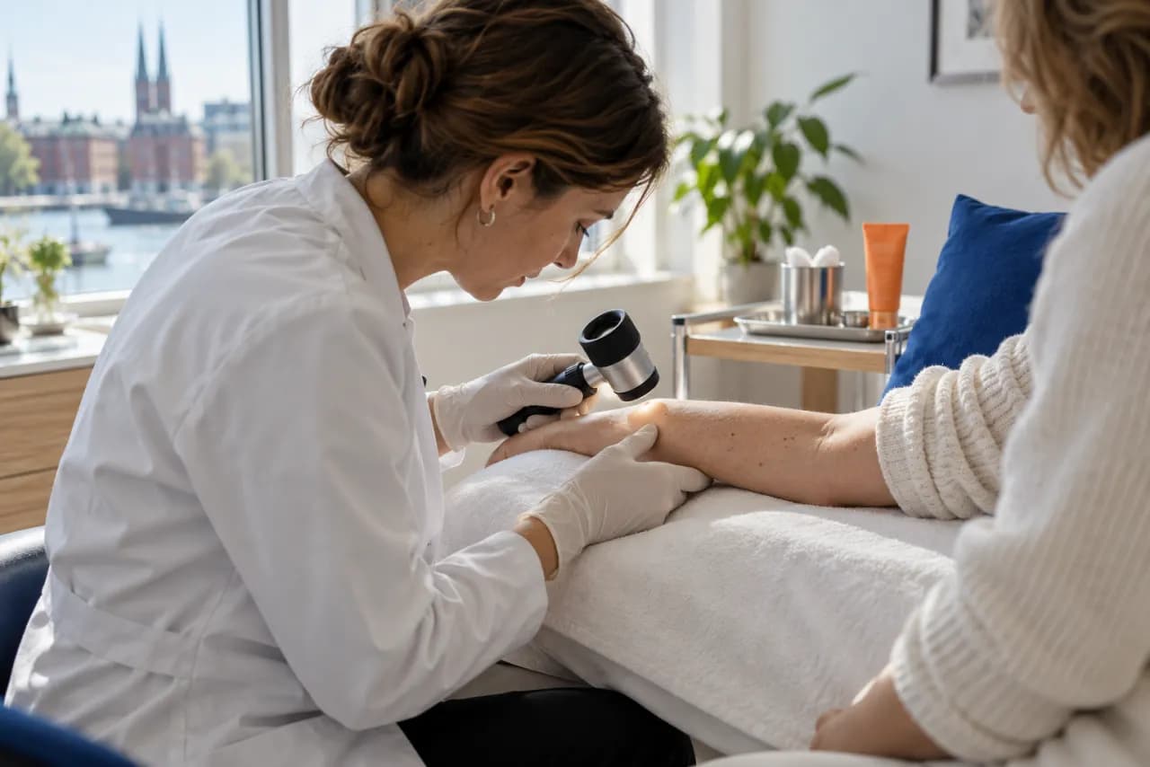
A patient uploads three dermoscopy photos of a changing mole before the clinic even opens. TUCAN triages the images into a ranked differential, drafts a structured consult note the dermatologist edits in seconds, and attaches the matching GOÄ code — so by the first coffee the case is reviewed, documented, and billable.

What your practice gains

Faster triage

Lesions are ranked and surfaced before the clinician opens the chart.

Change caught early

Serial images are compared against prior visits so evolving lesions are flagged.

Notes that keep pace

Visit documentation is drafted from the exam and imaging instead of typed after the clinic.

Coding without the afterthought

The matching GOÄ/EBM code is proposed with each note, so nothing billable slips away.

Follow-up that carries itself

Screening intervals and reviews are scheduled with prior images carried forward automatically.

Clinical Use Cases

Skin Cancer Screening

Systematic full-body skin examinations with AI-assisted mole mapping and risk assessment for early detection of melanoma.

Chronic Condition Management

Track treatment efficacy for conditions like psoriasis and eczema with standardized severity scoring and photo comparisons.

Cosmetic Consultations

Before-and-after documentation for aesthetic procedures with patient-facing galleries and outcome tracking.

What changes for your practice

Lesion intelligence

AI reads every lesion image the moment it arrives and hands the clinician a ranked, evidence-linked differential.

  • Ranked differential with confidence and reference imagery
  • Body-mapped lesion tracking across visits with automated measurements
  • Melanoma-risk flags surfaced before the appointment starts

Documentation that writes itself

Consult notes, photo consent, and coding are drafted from the encounter instead of typed after it.

  • Structured consult note generated from the image review
  • GDPR-compliant photo-consent capture attached to the record
  • One-click correction rather than blank-page dictation

Continuity of care

Follow-ups, recalls, and remote checks keep chronic and post-procedure patients on track without manual chasing.

  • Automated recall scheduling for high-risk skin-cancer screens
  • Side-by-side photo comparison for psoriasis and eczema tracking
  • Remote follow-up consults with the full lesion history in view

The connected pipeline

  1. Image capture

    The patient or clinic captures dermoscopy and clinical photos with standardized lighting and positioning templates.

  2. AI differential

    TUCAN analyzes the images and returns a ranked differential with confidence scores and comparable reference cases.

  3. Clinician review

    The dermatologist confirms or adjusts the AI findings and records the diagnosis directly in the chart.

  4. Structured note + coding

    A structured consult note is generated and the matching GOÄ/EBM billing code is attached automatically.

  5. Follow-up scheduling

    Recalls, remote skin checks, and treatment-tracking visits are scheduled with the lesion history carried forward.

How the data moves

Image and history capture

Dermoscopic images and history are captured through structured, mapped forms.

TUCAN lesion analysis

Images are read into a differential with change-over-time and risk signals.

FHIR record

Lesion maps, imaging, and findings are written to the patient's structured clinical record.

Structured note + coding

A visit note is drafted from the reviewed encounter for the dermatologist to confirm.

Surveillance schedule

Follow-up imaging and reviews are scheduled with prior lesion maps carried forward.

Where the AI works

Each agent owns a distinct part of the dermatology workflow, so the clinician stays in control of every decision.

TUCAN

Reads dermoscopic images into a differential and flags lesions that have changed since the last visit before the clinician opens the chart.

Condor

Drafts the structured visit note and lesion map from the reviewed encounter for one-click correction.

FlowAgent

Guides imaging intake and surveillance steps, keeping screening intervals and reviews on track.

TW
Thomas WeberMRN-2026-0847
Penicillin Allergy
Allergies
PenicillinAnaphylaxisSevere
LatexSkin RashModerate
IbuprofenNauseaMild
Medications
Metformin 1000mgTwice daily
Lisinopril 10mgOnce daily
Aspirin 100mgOnce daily
Recent Labs
HbA1c6.8%High
LDL Cholesterol98 mg/dLNormal
eGFR> 90Normal

How Dudoxx delivers it

AI lesion triage

Dermoscopy intake forms

Remote skin checks

GOÄ/EBM coding

Integrations

DICOM Imaging

Native DICOM support for dermoscopy images with PACS integration and standardized image storage.

Lab Connectivity

Direct lab order placement for biopsies and pathology with automated result retrieval and report linking.

Telemedicine

Secure image sharing and video consultations for remote dermatology assessments and follow-up appointments.

Ready to Transform Your Practice?

Get a personalized demo tailored to your specialty and practice size. All solutions are FHIR R4 native and run fully on-premise.